
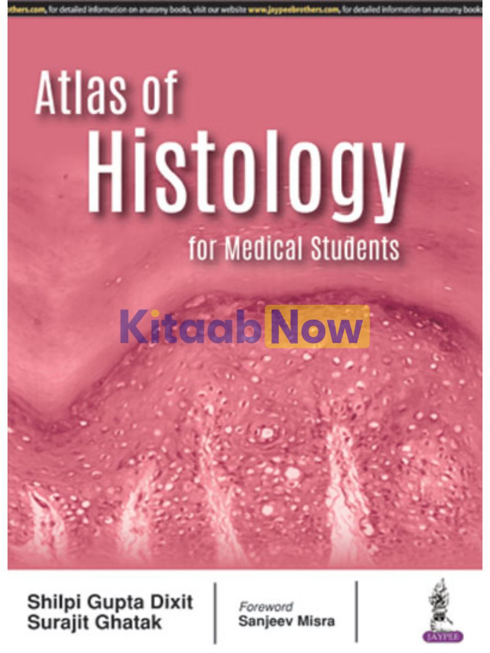
Atlas of Histology for Medical Students
Rs. 1,520
- Authors: Shilpi Gupta Dixit, Surajit Ghatak
- ISBN: 9789352701285
- Publisher: Jaypee Brothers Medical Publishers
- Edition: 1st
- Publication Date: August 15, 2017
- Format: Paperback – 102 pages
- Language: English
Out of stock
Share
Description
An atlas containing microphotographs of human tissue accompanied by hand-drawn diagrams for easy reproducibility. Each plate is well labelled and supplemented by salient identification features unique to the tissue or the organ.
Organized to maximize ease of use and speed of learning.
Subdivided into 19 major chapters that correspond to 19 major areas of study starting with cells, moving into tissues, and progressing into organ systems.
Of immense value to undergraduate and postgraduate students of anatomy and pathology.
Also helpful for students of paramedical and general science where histology is a part of the curriculum.
Table of Contents
- Parts of Light Microscope
- Electron Microscopic Structure of Cell
- Epithelium
- Glands
- Connective Tissue
- Cartilage
- Bone
- Muscle
- Nervous Tissue
- Cardiovascular System
- Lymphatic Tissue
- Integumentary System
- Digestive System
- Respiratory System
- Urinary System
- Male Reproductive System
- Female Reproductive System
- Endocrine System
- Special Senses
Additional information
| Weight | 0.345 kg |
|---|
Only logged in customers who have purchased this product may leave a review.
Related Products


Reviews
There are no reviews yet.