
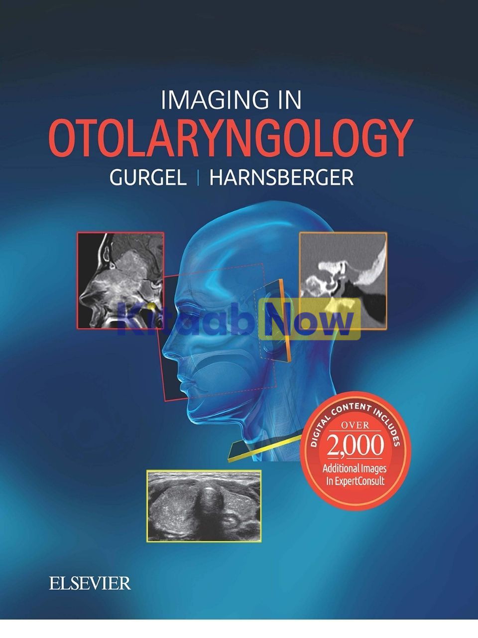
Imaging in Otolaryngology
- Authors: Richard Gurgel, H. Ric Harnsberger
- ISBN: 9780323545082
- Publisher: Elsevier Science Publishing
- Publication Date: April 11, 2018
- Format: Hardback – 525 pages
- Language: English
Description
Written directly to otolaryngologists, Imaging in Otolaryngology is a practical, superbly illustrated reference designed to enhance image reading skills at the point of care. Using annotated radiologic images, this unique reference provides the tools to quickly master the key points of imaging, all tailored to the needs of today’s otolaryngologist. Each one- or two-page chapter includes Key Facts and images, critical clinical information, definitions and clarifications of unfamiliar radiology jargon, and annotations.
Key Features
- Helps otolaryngologists at all levels of expertise – practicing clinicians, fellows, and residents – understand the significance of a given radiologic finding and next steps for the appropriate patient care (such as subsequent studies to order)
- Offers information in a quick-reference format that includes Key Facts, bulleted lists, and concise prose introductions for quick mastery of the material
- Provides comprehensive coverage accessible to residents and fellows, as well as an abundance of teaching pearls for the most experienced otolaryngologist
New to this Edition
- Contains 1,800 high-quality images relevant to otolaryngologists including radiology images, medical drawings, histopathology, gross pathology, and clinical photos
- Includes over 2,000 additional eBook images rich with other imaging modalities (MR, CT, ultrasound), clinical images, and histopathologic images
- Expert Consult™ eBook version included with purchase, which allows you to search all of the text, figures, and references from the book on a variety of devices
Table of Contents
- SECTION 1: Suprahyoid and Infrahyoid Neck
- Introduction and Overview
- Suprahyoid and Infrahyoid Neck Overview
- Parapharyngeal Space
- Parapharyngeal Space Overview
- Benign Tumors
- Parapharyngeal Space Benign Mixed Tumor
- Pharyngeal Mucosal Space
- Pharynx Overview
- Congenital Lesions
- Tornwaldt Cyst
- Infectious and Inflammatory Lesions
- Retention Cyst of Pharyngeal Mucosal Space
- Tonsillar Inflammation
- Tonsillar/Peritonsillar Abscess
- Benign and Malignant Tumors
- Minor Salivary Gland Malignancy of Pharyngeal Surface
- Non-Hodgkin Lymphoma of Pharyngeal Mucosal Space
- Masticator Space
- Masticator Space Overview
- Pseudolesions
- Pterygoid Venous Plexus Asymmetry
- Benign Masticator Muscle Hypertrophy
- CNV3 Motor Denervation
- Infectious Lesions
- Masticator Space Abscess
- Benign Tumors
- Masticator Space CNV3 Schwannoma
- Malignant Tumors
- Masticator Space CNV3 Perineural Tumor
- Masticator Space Chondrosarcoma
- Masticator Space Sarcoma
- Parotid Space
- Parotid Space Overview
- Infectious and Inflammatory Lesions
- Acute Parotitis
- Parotid Sjogren Syndrome
- Benign Lymphoepithelial Lesions-HIV
- Benign Tumors
- Parotid Benign Mixed Tumor
- Warthin Tumor
- Malignant Tumors
- Parotid Mucoepidermoid Carcinoma
- Parotid Adenoid Cystic Carcinoma
- Parotid Non-Hodgkin Lymphoma
- Metastatic Disease of Parotid Nodes
- Carotid Space
- Carotid Space Overview
- Normal Variants
- Tortuous Carotid Artery in Neck
- Vascular Lesions
- Carotid Artery Dissection in Neck
- Carotid Artery Pseudoaneurysm in Neck
- Carotid Artery Fibromuscular Dysplasia in Neck
- Acute Idiopathic Carotidynia
- Jugular Vein Thrombosis
- Post-Pharyngitis Venous Thrombosis (Lemierre)
- Benign Tumors
- Carotid Body Paraganglioma
- Glomus Vagale Paraganglioma
- Carotid Space Schwannoma
- Sympathetic Schwannoma
- Carotid Space Neurofibroma
- Retropharyngeal Space
- Retropharyngeal Space Overview
- Infectious and Inflammatory Lesions
- Reactive Adenopathy of Retropharyngeal Space
- Suppurative Adenopathy of Retropharyngeal Space
- Retropharyngeal Space Abscess
- Retropharyngeal Space Edema
- Metastatic Tumors
- Nodal Squamous Cell Carcinoma of Retropharyngeal Space
- Nodal Non-Hodgkin Lymphoma in Retropharyngeal Space
- Perivertebral Space
- Perivertebral Space Overview
- Pseudolesions
- Infectious and Inflammatory Lesions
- Acute Calcific Longus Colli Tendonitis
- Perivertebral Space Infection
- Vascular Lesions
- Vertebral Artery Dissection in Neck
- Benign and Malignant Tumors
- Brachial Plexus Schwannoma in Perivertebral Space
- Chordoma in Perivertebral Space
- Vertebral Body Metastasis in Perivertebral Space
- Posterior Cervical Space
- Posterior Cervical Space Overview
- Benign Tumors
- Posterior Cervical Space Schwannoma
- Metastatic Tumors
- Squamous Cell Carcinoma in Spinal Accessory Node
- Visceral Space
- Visceral Space Overview
- Inflammatory Lesions
- Chronic Lymphocytic Thyroiditis (Hashimoto)
- Metabolic Disease
- Multinodular Goiter
- Benign Tumors
- Thyroid Adenoma
- Parathyroid Adenoma in Visceral Space
- Malignant Tumors
- Differentiated Thyroid Carcinoma
- Medullary Thyroid Carcinoma
- Anaplastic Thyroid Carcinoma
- Non-Hodgkin Lymphoma of Thyroid
- Thyroglossal Duct Cyst Carcinoma
- Cervical Esophageal Carcinoma
- Miscellaneous
- Esophagopharyngeal Diverticulum (Zenker)
- Colloid Cyst of Thyroid
- Lateral Cervical Esophageal Diverticulum
- Hypopharynx, Larynx, and Cervical Trachea
- Hypopharynx, Larynx, & Trachea Overview
- Infectious and Inflammatory Lesions
- Croup
- Epiglottitis in a Child
- Supraglottits
- Trauma
- Laryngeal Trauma
- Benign and Malignant Tumors
- Upper Airway Infantile Hemangioma
- Laryngeal Chondrosarcoma
- Treatment-related Lesions
- Post-Radiatio
- n Larynx
- Miscellaneous
- Laryngocele
- Vocal Cord Paralysis
- Acquired Subglottic-Tracheal Stenosis
- Lymph Nodes
- Lymph Node Overview
- Infectious and Inflammatory Lesions
- Reactive Lymph Nodes
- Suppurative Lymph Nodes
- Tuberculous Lymph Nodes
- Non-TB Mycobacterium Nodes
- Sarcoidosis Lymph Nodes
- Giant Lymph Node Hyperplasia (Castleman)
- Histiocytic Necrotizing Lymphadenitis (Kikuchi)
- Kimura Disease
- Malignant Tumors
- Nodal Non-Hodgkin Lymphoma in Neck
- Nodal Hodgkin Lymphoma in Neck
- Nodal Differentiated Thyroid Carcinoma
- Systemic Nodal Metastases in Neck
- Transspatial and Multispatial
- Transspatial & Multispatial Overview
- Normal Variants
- Prominent Thoracic Duct in Neck
- Benign Tumors
- Lipoma of Head and Neck
- Plexiform Neurofibroma of Head and Neck
- Malignant Tumors
- Posttransplantation Lymphoproliferative Disorder
- Non-Hodgkin Lymphoma of Head and Neck
- Miscellaneous
- Lymphocele of Neck
- Sinus Histiocytosis (Rosai-Dorfman) of Head and Neck
- Fibromatosis of Head and Neck
- Oral Cavity
- Oral Cavity Overview
- Pseudolesions
- Hypoglossal Nerve Motor Denervation
- Congenital Lesions
- Submandibular Space Accessory Salivary Tissue
- Oral Cavity Dermoid and Epidermoid
- Oral Cavity Lymphatic Malformation
- Lingual Thyroid
- Infectious and Inflammatory Lesions
- Ranula
- Oral Cavity Sialocele
- Submandibular Gland Sialadenitis
- Oral Cavity Abscess
- Benign Tumors
- Submandibular Gland Benign Mixed Tumor
- Palate Benign Mixed Tumor
- Malignant Tumors
- Sublingual Gland Carcinoma
- Submandibular Gland Carcinoma
- Oral Cavity Minor Salivary Gland Malignancy
- Submandibular Space Nodal Non-Hodgkin Lymphoma
- Submandibular Space Nodal Squamous Cell Carcinoma
- Mandible-Maxilla and Temporomandibular Joint
- Mandible-Maxilla and TMJ Overview
- Congenital Lesions
- Solitary Median Maxillary Central Incisor
- Nonneoplastic Cysts
- Nasolabial Cyst
- Periapical Cyst (Radicular)
- Dentigerous Cyst
- Simple Bone Cyst (Traumatic)
- Nasopalatine Duct Cyst
- Infectious and Inflammatory Lesions
- TMJ Juvenile Idiopathic Arthritis
- Mandible-Maxilla Osteomyelitis
- Tumor-like Lesions
- TMJ Calcium Pyrophosphate Dihydrate Deposition Disease
- TMJ Pigmented Villonodular Synovitis
- TMJ Synovial Chondromatosis
- Benign and Malignant Tumors
- Ameloblastoma
- Keratocystic Odontogenic Tumor (Odontogenic Keratocyst)
- Mandible-Maxilla Osteosarcoma
- Treatment-related Lesions
- Mandible-Maxilla Osteoradionecrosis
- Mandible-Maxilla Osteonecrosis
- SECTION 2: Squamous Cell Carcinoma
- Squamous Cell Carcinoma Introduction and Overview
- Primary Sites, Perineural Tumor and Nodes
- Nasopharyngeal Carcinoma
- Nasopharyngeal Carcinoma
- Oropharyngeal Carcinoma
- Base of Tongue Squamous Cell Carcinoma
- Palatine Tonsil Squamous Cell Carcinoma
- Posterior Oropharyngeal Wall Squamous Cell Carcinoma
- HPV-related Oropharyngeal Squamous Cell Carcinoma
- Oral Cavity Carcinoma
- Oral Tongue Squamous Cell Carcinoma
- Floor of Mouth Squamous Cell Carcinoma
- Alveolar Ridge Squamous Cell Carcinoma
- Retromolar Trigone Squamous Cell Carcinoma
- Buccal Mucosa Squamous Cell Carcinoma
- Hard Palate Squamous Cell Carcinoma
- Hypopharyngeal Carcinoma
- Pyriform Sinus Squamous Cell Carcinoma
- Postcricoid Region Squamous Cell Carcinoma
- Posterior Hypopharyngeal Wall Squamous Cell Carcinoma
- Laryngeal Carcinoma
- Supraglottic Laryngeal Squamous Cell Carcinoma
- Glottic Laryngeal Squamous Cell Carcinoma
- Subglottic Laryngeal Squamous Cell Carcinoma
- Laryngeal Squamous Cell Carcinoma With Secondary Laryngocele
- Perineural Tumor
- 172 Perineural Tumor Spread
- Squamous Cell Carcinoma Lymph Nodes
- Nodal Squamous Cell Carcinoma
- Post-Treatment Neck
- Nodal Dissection in Neck
- Reconstruction Flaps in Neck
- Expected Changes of Neck Radiation Therapy
- Complications of Neck Radiation Therapy
- Osteoradionecrosis
- Post Laryngectomy
- SECTION 3: Pediatric and Syndromic Diseases
- Pediatric Lesions
- Congenital Overview
- Congenital Lesions
- Lymphatic Malformation
- Venous Malformation
- Congenital Vallecular Cyst
- Cervical Thymic Cyst
- 1st Branchial Cleft Cyst
- 2nd Branchial Cleft Cyst
- 3rd Branchial Cleft Cyst
- 4th Branchial Cleft Cyst
- Dermoid and Epidermoid
- Trauma
- Fibromatosis Colli
- Benign Tumors
- Infantile Hemangioma
- Malignant Tumors
- Rhabdomyosarcoma
- Syndromic Diseases
- Neurofibromatosis Type 1
- Neurofibromatosis Type 2
- Basal Cell Nevus Syndrome
- Branchiootorenal Syndrome
- CHARGE Syndrome
- Hemifacial Microsomia
- Treacher Collins Syndrome
- Pierre Robin Sequence
- X-Linked Stapes Gusher (DFNX2)
- McCune-Albright Syndrome
- Cherubism
- SECTION 4: Sinonasal Cavities and Orbit
- Nose and Sinus
- Sinonasal Overview
- Congenital Lesions
- Nasolacrimal Duct Mucocele
- Choanal Atresia
- Nasal Glioma
- Nasal Dermal Sinus
- Frontoethmoidal Cephalocele
- Congenital Nasal Pyriform Aperture Stenosis
- Infectious and Inflammatory Lesions
- Acute Rhinosinusitis
- Chronic Rhinosinusitis
- Complications of Rhinosinusitis
- Allergic Fungal Sinusitis
- Sinus Mycetoma
- Invasive Fungal Sinusitis
- Sinonasal Polyposis
- Solitary Sinonasal Polyp
- Sinonasal Mucocele
- Silent Sinus Syndrome
- Granulomatosis With Polyangiitis (Wegener)
- Nasal Cocaine Necrosis
- Benign Tumors and Tumor-like Lesions
- Sinonasal Fibrous Dysplasia
- Sinonasal Osteoma
- Sinonasal Ossifying Fibroma
- Juvenile Angiofibroma
- Sinonasal Inverted Papilloma
- Sinonasal Hemangioma
- Malignant Tumors
- Sinonasal Squamous Cell Carcinoma
- Esthesioneuroblastoma
- Sinonasal Adenocarcinoma
- Sinonasal Melanoma
- Sinonasal Non-Hodgkin Lymphoma
- Sinonasal Undifferentiated Carcinoma
- Sinonasal Adenoid Cystic Carcinoma
- Sinonasal Chondrosarcoma
- Orbit
- Orbit Overview
- Congenital Lesions
- Coloboma
- Orbital Dermoid and Epidermoid
- Vascular Lesions
- Orbital Lymphatic Malformation
- Orbital Cavernous Venous Malformation (Hemangioma)
- Infectious and Inflammatory Lesions
- Orbital Subperiosteal Abscess
- Orbital Cellulitis
- Idiopathic Orbital Inflammatory (Pseudotumor)
- Thyroid-Associated Orbitopathy
- Optic Neuritis
- Benign Tumors
- Orbital Infantile Hemangioma
- Optic Nerve Sheath Meningioma
- Malignant Tumors
- Retinoblastoma
- SECTION 5: Skull Base
- Skull Base Lesions
- Skull Base Overview
- Clivus
- Ecchordosis Physaliphora
- Invasive Pituitary Macroadenoma
- Chordoma
- Sphenoid Bone
- Persistent Craniopharyngeal Canal
- Sphenoid Benign Fatty Lesion
- Central Skull Base Trigeminal Schwannoma
- Occipital Bone
- Hypoglossal Nerve Schwannoma
- Jugular Foramen
- Jugular Bulb Pseudolesion
- High Jugular Bulb
- Dehiscent Jugular Bulb
- Jugular Bulb Diverticulum
- Glomus Jugulare Paraganglioma
- Jugular Foramen Schwannoma
- Jugular Foramen Meningioma
- Dural Sinuses
- Dural Sinus and Aberrant Arachnoid Granulations
- Skull Base Dural Sinus Thrombosis
- Cavernous Sinus Thrombosis
- Dural Arteriovenous Fistula
- Diffuse or Multifocal Skull Base Disease
- Skull Base Cephalocele
- Skull Base CSF Leak
- Skull Base Fibrous Dysplasia
- Skull Base Paget Disease
- Skull Base Langerhans Cell Histiocytosis
- Skull Base Osteopetrosis
- Skull Base Idiopathic Inflammatory Pseudotumor
- Skull Base Giant Cell Tumor
- Skull Base Meningioma
- Skull Base Plasmacytoma
- Skull Base Multiple Myeloma
- Skull Base Metastasis
- Skull Base Chondrosarcoma
- Skull Base Osteosarcoma
- Skull Base and Facial Trauma
- Skull Base and Facial Trauma Overview
- Skull Base
- Temporal Bone Fractures
- Ossicular Dislocations and Disruptions
- Skull Base Trauma
- Facial Bones
- Orbital Foreign Body
- Orbital Blowout Fracture
- Transfacial Fractures (Le Fort)
- Zygomaticomaxillary Complex Fracture
- Complex Facial Fracture
- Nasoorbitalethmoidal Fracture
- Mandible Fracture
- SECTION 6: Temporal Bone and CPA-IAC
- Introduction and Overview
- Temporal Bone Overview
- External Auditory Canal
- Congenital Lesions
- Foramen Tympanicum
Author Biography
Richard K. Gurgel, MD, Assistant Professor Otolaryngology – Head and Neck Surgery, University of Utah School of Medicine, Salt Lake City, Utah and
H. Ric Harnsberger, MD, R. C. Willey Chair in Neuroradiology, Professor of Radiology & Otolaryngology, University of Utah School of Medicine, Salt Lake City, Utah
Additional information
| Weight | 1.900 kg |
|---|
Only logged in customers who have purchased this product may leave a review.
Related Products




Reviews
There are no reviews yet.