
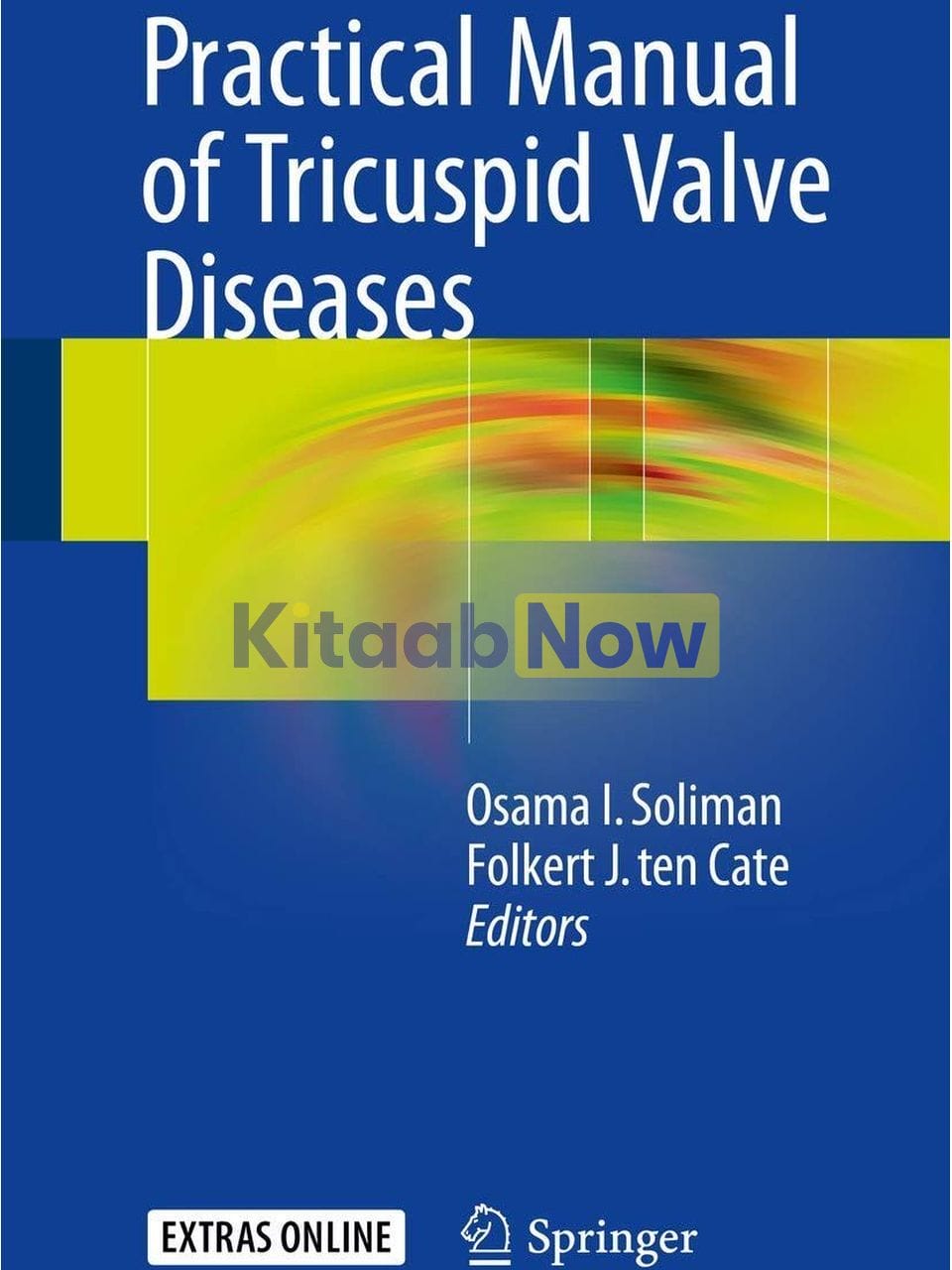
Practical Manual of Tricuspid Valve Diseases
- Edited by: Osama I. Soliman, Folkert J. ten Cate
- ISBN: 9783319582283
- Publisher: Springer Publishing Company
- Publication Date: October 23, 2017
- Format: Paperback – 412 pages
- Language: English
Description
This book provides a comprehensive overview of tricuspid valve disease and an overview of different therapeutic options such as surgical and percutaneous approaches, current clinical trials and registries in the field of tricuspid valve disease. The book has an educational goal with key messages highlighted at the end of each chapter to assist comprehension of the often complex concepts. Practical Manual of Tricuspid Valve Diseases assists the clinician in dealing with different manifestations of tricuspid valve disease and providing help for choosing the best option for the management of these patients. It therefore serves as the standard reference regarding all aspects of tricuspid valve disease in adults and will be of importance to all medical professionals involved in the management of these patients.
Table of Contents
- Part I: Anatomical and Pathological Perspective
- Chapter 1: Anatomy and Pathology of the Tricuspid Valve
- Components of the Tricuspid Valve
- The Tricuspid Leaflets and Commissures
- Tendinous Cords and Papillary Muscles
- Pathological Changes in the Tricuspid Valve
- The Tricuspid Valve in Disease
- Tricuspid Regurgitation
- Valvar Stenosis
- Straddling Tricuspid Valve
- Conclusion
- Take Home Points
- Review Questions
- References
- Part II: Clinical Perspective
- Chapter 2: Clinical Recognition of Tricuspid Valve Disease
- Tricuspid Stenosis
- Frequency [2]
- Natural History
- Clinical Features
- Common Symptoms
- Less Common Symptoms
- Rare Symptoms
- Aetiology and Pathogenesis
- Rheumatic Heart Disease
- Congenital Tricuspid Stenosis
- Infective Endocarditis
- Carcinoid Heart Disease
- Unusual Causes
- Differential Diagnosis
- Investigations
- Management
- Consultations
- Tricuspid Regurgitation
- Pathophysiology
- Physical Examination
- Differential Diagnosis
- Investigations
- Natural History and Prognosis
- Medical Therapy
- Long-Term Monitoring
- Approach Considerations
- Specific Conditions
- Tricuspid Atresia
- Presentation
- Investigations
- Management
- Complications
- Prognosis
- Review Questions
- Select the Single Best Answer
- Case Studies
- References
- Chapter 3: Tricuspid Regurgitation in Patients with Heart Transplant
- Introduction
- Case
- Epidemiology
- Pathophysiology
- Clinical Features
- Management and Prognosis
- Key Points
- Review Questions
- Select the Single Best Sentence
- References
- Chapter 4: Tricuspid Regurgitation in Patients with Pacemakers and Implantable Cardiac Defibrilla
- Introduction
- Pathophysiology and Mechanisms (See Fig. 4.1)
- Clinical Presentation
- Imaging
- Management (See Fig. 4.4)
- Prevention (See Table 4.2)
- Summary
- Review Questions
- References
- Part III: Non-invasive Imaging Perspective
- Chapter 5: Tricuspid Valve Disease: Imaging Using Transthoracic Echocardiography
- Introduction
- Tricuspid Valve Apparatus
- TTE Examination of the Tricuspid Valve
- TTE Modes
- 2D–TTE
- 3D-TTE
- Acquisition
- Analysis and Display
- Incremental Value of 3D-TTE over 2D-TTE of the TV
- Simultaneous Multiplane Imaging
- Bi-plane TTE
- iRotate Mode
- TTE Approach to Tricuspid Valve Disease
- TTE Approach to Tricuspid Valve Stenosis
- TTE Approach to Tricuspid Valve Regurgitation
- TTE Diagnostic Algorithm of TR
- Etiology and Mechanisms of TR
- Grading of TR Severity
- Use of Colour Doppler in TR Severity Assessment
- TR Jet Area
- TR Jet Vena Contracta
- Flow convergence method (proximal isovelocity surface area [PISA] method). Flow Acceleration and T
- Method 1. PISA Quantification of TR
- Method 2. PISA Quantification of TR
- Caveats in PISA Estimation of TR
- PISA Quantification on 3DE
- TR Jet on Continuous-Wave Doppler
- Hepatic Vein Flow in Patients with TR
- Other Surrogate Signs of TR Severity
- Assessment of TR Severity in Functional TR (Special Considerations)
- TTE Assessment of the Tricuspid Valve Annulus
- Key Points
- Review Questions
- Select the Single Best Sentence
- References
- Chapter 6: Imaging of the Tricuspid Valve: Transoesophageal Echocardiography
- Introduction
- TOE Imaging for Tricuspid Valve Imaging
- Grading Severity of Tricuspid Regurgitation
- Review Questions
- Select the Single Best Sentence
- References
- Chapter 7: A Surgeon’s View on Echocardiographic Imaging of the Tricuspid Valve
- Introduction
- Tricuspid Annulus
- Degree of Tricuspid Regurgitation
- Morphology
- Tethering
- Case 1: Functional Tricuspid Regurgitation with a Structural Component
- Heart Team Evaluation
- Operation
- Postoperative Period
- Case 2: Cardiac Endovascular Pacemaker-Lead Interference of the Tricuspid Valve
- Heart Team Evaluation
- Operation
- Postoperative Period
- Case 3: Functional Regurgitation Due to Left Sided Heart Disease
- Heart Team Evaluation
- Operation
- Postoperative Period
- Discussion
- Take Home Message
- Review Questions
- References
- Chapter 8: Imaging of the Tricuspid Valve: Magnetic Resonance Imaging
- Introduction
- TV Anatomy
- Comprehensive Evaluation of Tricuspid Valve by CMR
- Assessment of Tricuspid Valve Anatomy
- CMR Sequence and Image Acquisition
- Advantages
- Limitations
- Assessment of Tricuspid Valve Flow
- CMR Sequence and Image Acquisition
- Assessment of Right Ventricle Volume and Function
- CMR Sequence and Image Acquisition
- Assessment of Myocardial Fibrosis
- CMR Sequence and Image Acquisition
- Limitations
- Classification of TV Dysfunction
- Tricuspid Valve Regurgitation
- Secondary Tricuspid Valve Regurgitation
- Primary Tricuspid Valve Regurgitation
- Tricuspid Valve Stenosis
- CMR Imaging for Secondary Tricuspid Valve Regurgitation
- CMR Imaging for Primary Tricuspid Valve Regurgitation
- Ebstein’s Anomaly
- Rheumatic Heart Disease
- Infective Endocarditis
- Carcinoid Syndrome
- Tumors
- Traumatic
- CMR Imaging for Tricuspid Stenosis (TS)
- Conclusion
- Review Questions
- Select the Single Best Sentence
- References
- Chapter 9: Tricuspid Valve Disease: A Computed Tomographic Assessment
- Imaging Technics of the Tricuspid Valve Using CT
- Scan Mode
- Contrast Injection
- Clinical Utility of Computed Tomography in Right Heart Diseases
- Computed Tomography Acquisition Techniques for the Right Side of the Heart
- Clinical Utility of Cardiac CT
- Comprehensive Assessment of Tricuspid Valve and Surrounding Structures by CT
- The Tricuspid Valve
- Tricuspid Annulus
- CT Assessment of the Tricuspid Valve and Adjacent Anatomical Structures
- Advantages
- Limitations
- The Right Coronary Artery
- The Right Ventricular Apex
- The Vena Cava
- Surgical Interventions for the Tricuspid Valve
- Clinical Utility of CT in Planning Transcatheter Interventions for Functional TR
- Percutaneous Tricuspid Annuloplasty
- Tricuspid Orifice
- Caval Valve Implantation Concept
- Conclusion
- Appendix
- Review Questions
- Select the Single Best Sentence
- References
- Chapter 10: Tricuspid Annulus Measurements: Dynamic Changes in Health and Disease
- Introduction
- Normal Tricuspid Annulus: Size, Shape and Dynamics
- Changes in Tricuspid Annulus Geometry and Function in Functional Tricuspid Regurgitation
- Measurement of Tricuspid Annulus Dilation
- Tricuspid Annulus Deformation: A “New” Pathophysiologic Entity for TR Development?
- Unmet Needs and Future Research Directions
- Conclusions
- Review Questions
- Select the Single Best Sentence
- References
- Chapter 11: Imaging of the Right Ventricle: Overview of Imaging Modalities for Assessing RV Volu
- Introduction
- Right Ventricular Anatomy and Physiology
- Imaging Modalities for Assessment of RV Function and Size, and Tricuspid Annulus Size
- Echocardiography
- RV Function
- Right Ventricular Size
- Cardiovascular Magnetic Resonance
- Multidetector Computed Tomography (MDCT)
- Radionuclide Techniques
- Summary
- References
- Part IV: Hemodynamic Perspective
- Chapter 12: Echocardiographic Assessment of Pulmonary Artery Pressure, Tips and Tricks
- Introduction
- The Anatomy of the Right Ventricle and the Effects of Elevated Pulmonary Arterial Pressures
- Parasternal Long Axis View of the Left Ventricle
- Parasternal Long Axis of the Right Ventricle
- Parasternal Long and Short Axis Views of the Pulmonary Valve and Main Pulmonary Artery
- Parasternal Short Axis at the Level of the Papillary Muscles
- Apical 4 Chamber View
- Tricuspid Valve Anatomy and Effect of High Pulmonary Artery Pressures and Right Ventricle Dilata
- Pulmonary Pressure Estimation
- Pulmonary Artery Pressure Estimation Using Tricuspid Valve Regurgitation
- Pulmonary Artery Pressure Estimation Using Pulmonary Valve Regurgitation
- Pulmonary Artery Pressure Estimation Using the Right Ventricle Outflow Pulsed Wave Doppler Velocit
- Pulmonary Artery Diameter
- Questions
- Review Questions
- References
- Chapter 13: Right Heart Catheterization for Hemodynamic Evaluation of Right Sided Heart Disease
- Introduction
- Basics of the Right Heart Catheterization
- Tips and Tricks
- Hemodynamic Information from a RHC
- Calculation of Cardiac Output
- Assessment and Calculation of Intra-Cardiac Shunts
- Resistance of the Vascular Bed
- Conclusion
- Review Questions
- References
- Part V: Therapeutic Perspective
- Chapter 14: Pharmacotherapy of Tricuspid Valve Disease
- Introduction
- Case
- Pharmacotherapy (See Fig. 14.3)
- Lifestyle Advises
- Loop Diuretics
- Mineralocorticoid Receptor Antagonist
- Digoxin
- ACE-Inhibitors, Angiotensin II Receptor Blocker and Beta-Blockers
- References
- Chapter 15: Tricuspid Valve Interventions: Heart Team Discussion, When, Who and What?
- Prevalence and Mechanism of Tricuspid Regurgitation
- Outcomes
- Surgery for TR
- Transcatheter Interventions for TR
- Imaging of the Tricuspid Valve
- Transthoracic Echocardiography
- Transesophageal Echocardiography
- Transcatheter Treatment Modalities
- Bicaval Valve Implantation
- Devices to Improve Leaflet Apposition
- Annuloplasty Devices
- Preclinical Experience with Upcoming Devices
- Transcatheter Valve Replacement
- Conclusions
- References
- Chapter 16: Tricuspid Valve Disease: Surgical Outcome
- Introduction
- Tricuspid valve Repair and Replacement
- Tricuspid Valve Repair
- Patient Presentation and Intraoperative Details
- Outcomes
- Tricuspid Valve Replacement
- Patient Presentation and Intraoperative Details
- Outcome
- Outcomes of Bioprosthesis vs. Mechanical Prosthesis
- Functional Tricuspid Regurgitation
- Patient Presentation and Intraoperative Details
- Outcome
- Rheumatic Tricuspid Valve Disease
- Patient Presentation and Intraoperative Details
- Outcomes
- Congenital Tricuspid Valve Disease
- Patient Presentation and Intraoperative Details
- Outcome
- Carcinoid Disease of the Tricuspid Valve
- Iatrogenic Damage of the Tricuspid Valve
- Endocarditis of the Tricuspid Valve
- Take Home Message
- Review Questions
- References
- Chapter 17: Tricuspid Valve Disease: Surgical Techniques
- Preparation for Cardio-Pulmonary Bypass
- Access
- Cannulation
- Exposure
- Surgical Anatomy and Analysis
- Tricuspid Valve Repair for Functional Disease
- Suture Annuloplasty
- Kay Procedure
- De Vega Procedure
- Ring Annuloplasty
- Enlargement of the Anterior Leaflet
- “Clover Technique”
- Results
- Tricuspid Valve Repair for Primary Disease
- Intervention on the Leaflets
- Intervention on the Chordae
- Intervention on the Papillary Muscles
- Intervention on the Commissures
- Tricuspid Valve Replacement
- Choice of the Prosthetic Valve
- Surgical Technique
- Results
- Special Condition
- Tricuspid Valve Endocarditis
- Ebstein’s Anomaly
- Carcinoid Disease
- Traumatic Lesion
- Pacemaker Lead-Induced Tricuspid Regurgitation
- Late Tricuspid Regurgitation Following Previous Left-Sided Valve Surgery
- Case Report
- References
- Chapter 18: Transcatheter Interventions for Tricuspid Regurgitation: Rationale, Overview of Curren
- Rationale for Transcatheter Interventions for Tricuspid Regurgitation
- Anatomical Challenges to Transcatheter Tricuspid Valve Interventions
- Approaches for Transcatheter Tricuspid Valve Interventions
- Orthotopic Valve Implantation
- Native Tricuspid Valve Replacement
- Dedicated Transcatheter TV Bioprosthesis
- Non-Dedicated Transcatheter Heart Valve Implantation in the Tricuspid Position
- Transcatheter Valve in Malfunctioning Surgical Bioprosthetic Valve
- Transcatheter Valve in Tricuspid Repair Ring
- Heterotopic (Vena Caval) Valve Implantation
- Self-Expanding (Dedicated) Valve
- Balloon-Expandable Transcatheter Heart Valves
- Transcatheter Tricuspid Valve Repair
- Annular Reduction/Remodeling Devices
- The Mitralign/Trialign Device
- The TriCinch System
- The Transatrial Intrapericardial Tricuspid Annuloplasty (TRAIPTA) Device
- Surgical-Like Annuloplasty Rings
- Coaptation Devices
- The FORMA Repair System and the TV Occluder Device
- The MitraClip Device
- Open Issues and Future Perspective
- Safety Issues
- Preprocedural Planning and Procedural Guidance
- What Efficacy Endpoints are Suitable for TTVIs Studies?
- References
- Chapter 19: Catheter-Based Therapy for Tricuspid Valve Disease: Practical Considerations for Interve
- Introduction
- Patients’ Selection: Which Patient Could Benefit the Most from Interventional Tricuspid Therapi
- Anatomical Considerations
- The Importance of Imaging: Patients’ Eligibility and Intraprocedural Guidance
- Specific Considerations for Transcatheter Tricuspid Valve in Valve and Valve in Ring
- Specific Considerations for Transcatheter Tricuspid Valve Repair Devices
- Tricuspid Clipping
- Tricuspid Annuloplasy Devices
- Caval Valve Implantation
- Pericardial Device
- Tricuspid Valve Spacer
- How to Report the Outcome?
- Conclusions
- Review Questions for Chapters 15, 18 and 19
- Select the Single Best Sentence
- Answers
- References
- Index
Editors Biography
Dr Osama I. Soliman, MD, PhD, FACC, FESC works as assistant professor of cardiology at the Thoraxcenter, Erasmus MC: University Medical Center Rotterdam, Rotterdam, the Netherlands. He is the director of structural heart imaging at Cardialysis, Rotterdam, The Netherlands. Dr Soliman is a consultant cardiologist specialised in non-invasive cardiovascular imaging and he is a principal investigator of several clinical trials.
Dr Soliman has been graduated in 1996 Summa Cum Laude from the Medical School of Al-Azhar University, Cairo, Egypt. He completed eight years of training in internal medicine and cardiology at Al-Azhar University Hospitals in Cairo, Egypt. In 2000, he was awarded the degree of Master of Science Cum Laude based on a dissertation entitled “Intravascular ultrasound versus quantitative coronary angiography for the assessment of immediate results of coronary artery stenting”. In 2005, he joined the Thoraxcenter, Erasmus MC:University Medical Center Rotterdam, Rotterdam, the Netherlands where he was granted the doctoral degree from the Erasmus University Rotterdam, the Netherlands in 2007 based on dissertation entitled “Advanced quantitative echocardiography guiding therapy for heart failure”.
His major areas of teaching and research include echocardiography and non-invasive imaging and heart failure. He is leading a research team in structural heart interventions and outcome studies. His bibliography to date includes more than 200 articles and book chapters.
Dr. Soliman is an active member of several professional societies and organizations including the American College of Cardiology, the European Society of Cardiology, the American Society of Echocardiography, the International Society of Contrast Ultrasound. He is a member of the Editorial Board of several prestigious cardiology journals including the Journal of the American Society of Echocardiography and Eurointervention.
Dr Folkert J. ten Cate MD, PhD, FESC, FACC is an Associate Professor of the Department of Cardiology at the Thoraxcenter, Erasmus MC: University Medical Center Rotterdam, Rotterdam, the Netherlands. He has been the late director of the Thoraxcenter fellow training program. Dr ten Cate is a consultant cardiologist specialised in general cardiology and non-invasive cardiovascular imaging.
Dr ten Cate has been graduated in 1972 from the Medical School of Erasmus University, Rotterdam, The Netherlands. He completed six years of training in internal medicine and cardiology at Erasmus University Hospitals in Rotterdam, The Netherlands. In 1973, he joined the Erasmus MC: University Medical Center Rotterdam, Rotterdam, the Netherlands, where he was granted the doctoral degree from the Erasmus University Rotterdam in 1978 based on a dissertation entitled “Asymmetric septal hypertrophy (ASH): echocardiographic manifestations dissertation”.
He has given numerous international and national lectures. He is a world renown expert in Hypertrophic Cardiomyopathy and Contrast Echocardiography. Also he has trained numerous foreign and national fellows in cardiology.
His major areas of teaching and research include echocardiography and non-invasive imaging, cardiomyopathies, valvular heart disease, and diastolic heart failure. His bibliography to date includes more than 350 articles and book chapters.
Dr. ten Cate is an active member of several professional societies and organizations including the American College of Cardiology, the European Society of Cardiology and the International Society of Contrast Ultrasound.
Additional information
| Weight | 0.658 kg |
|---|
Only logged in customers who have purchased this product may leave a review.
Related Products




Reviews
There are no reviews yet.