
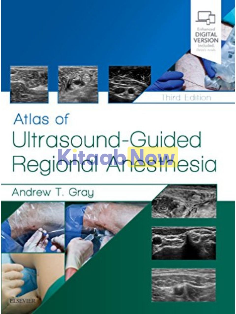
Atlas of Ultrasound-Guided Regional Anesthesia (3rd Edition)
Rs. 70,600
- Authors: Andrew T. Gray
- ISBN: 9780323509510
- Publisher: Elsevier Science Publishing
- Edition: 3rd
- Publication Date: May 09, 2018
- Format: Paperback – 412 pages
- Language: English
Share
Description
Step-by-step videos and images, board-style review questions, and coverage of new blocks make this highly respected title a must-have reference for clinical practice. Written by Andrew T. Gray, MD, PhD, one of the pioneers of the use of ultrasound to guide needle placement, Atlas of Ultrasound-Guided Regional Anesthesia, 3rd Edition, shows you how to safely and effectively use the latest methods and applications of this technique.
Key Features
- Board-style review questions found on the Expert Consult eBook version test your knowledge and help you prepare for the ABA exam.
- Helps ensure correct needle placement with numerous 3-D and long-axis views that clearly depict surrounding structures.
New to this Edition
- Includes coverage of 11 new blocks: Adductor Canal, Posterior Femoral Cutaneous, Pectoral, Quadratus Lumborum, Pudendal, Paravertebral, Transversus thoracis, Supraorbital, Transtracheal, Greater Occipital and Lesser Occipital.
- Features access to 20 author-narrated videos showing proper placement of needles using ultrasound guidance, including 11 new videos: Forearm (ulnar, median and radial), Ankle (tibial, saphenous, superficial peroneal, deep peroneal, sural), Paravertebral, Adductor Canal, and Catheters.
- Presents several new chapters, including Regional Anesthesia in Resource-Constrained Environments and Safety of Ultrasound Guided Regional Blocks.
- Expert Consult™ eBook version included with purchase. This enhanced eBook experience allows you to search all of the text, figures, and references from the book on a variety of devices.
Table of Contents
- Section 1 – Introduction to Ultrasound Imaging
- Ultrasound
- Speed of Sound
- Attenuation
- Reflection
- Beam Width (Slice Thickness)
- Anisotropy
- Spatial Compound Imaging
- Doppler Imaging
- Ultrasound Transducers
- Transducer Manipulation
- Needle Imaging
- Approach and Techniques
- Sonographic Signs of Successful Injections
- Ultrasound-Guided Continuous Peripheral Nerve Blocks
- Three-Dimensional Ultrasound
- Section 2 – Structures
- Anatomic Structures
- Skin and Subcutaneous Tissue
- Peripheral Nerves
- Muscle
- Tendons and Ligaments
- Arteries
- Veins
- Bone
- Pleura
- Peritoneum
- Section 3 – Upper Extremity Blocks
- Lymph Nodes
- Supraclavicular Nerve Block
- Interscalene and Supraclavicular Blocks
- Phrenic Nerve Imaging
- Dorsal Scapular Nerve Imaging
- Suprascapular Nerve Block (Anterior Approach above the Clavicle)
- Infraclavicular Block
- Axillary Block
- Musculocutaneous Nerve Block
- Forearm Blocks
- Radial Nerve Block
- Median Nerve Block
- Ulnar Nerve Block
- Section 4 – Lower Extremity Blocks
- Lateral Femoral Cutaneous Nerve Block
- Fascia Iliaca Block
- Femoral Nerve Block
- The Adductor Canal Block
- Obturator Nerve Block
- Sciatic Nerve Block
- Anterior Sciatic Nerve Block
- Popliteal Block
- Posterior Femoral Cutaneous Nerve (PFCN) Block
- Ankle Block
- Saphenous Nerve Block in the Leg
- Deep Peroneal Nerve Block
- Superficial Peroneal Nerve Block
- Sural Nerve Block
- Tibial Nerve Block
- Section 5 – Trunk Blocks
- Intercostal Nerve Block
- Pectoral Nerve Block (the Pecs Block)
- Transversus Thoracis Muscular Plane Block
- Rectus Sheath Block
- Ilioinguinal and Iliohypogastric Nerve Blocks
- Transversus Abdominis Plane (TAP) Blocks
- Quadratus Lumborum Blocks
- Thoracic Paravertebral Block
- Pudendal Nerve Block in Children
- Neuraxial Block
- Caudal Epidural Block
- Section 6 – Head and Neck Blocks
- Supraorbital Nerve Block
- Mental Nerve Block
- Superior Laryngeal Nerve Block
- Transtracheal Block
- Greater Occipital Nerve Block
- Lesser Occipital Nerve Block
- Great Auricular Nerve Block
- Cervical Plexus Block
- Stellate Ganglion Block (Cervicothoracic Sympathetic Ganglion Block)
- Section 7 – Safety Issues
- Safety: Practical Techniques to Prevent Complications During Ultrasound-Guided Nerve Blocks
- Regional Anesthesia in Resource-Constrained Environments
- Appendix 1. Self-Assessment Questions: Text
- Appendix 2. Self-Assessment Questions: Images
- Appendix 3. Advanced Self-Assessment Questions: Text
- Appendix 4. Advanced Self-Assessment Questions: Images
Author Biography
By Andrew T. Gray, MD, Department of Anesthesia and Perioperative Care, UCSF School of Medicine San Francisco, CA
Additional information
| Weight | 1.400 kg |
|---|
Only logged in customers who have purchased this product may leave a review.
Related Products





Reviews
There are no reviews yet.