
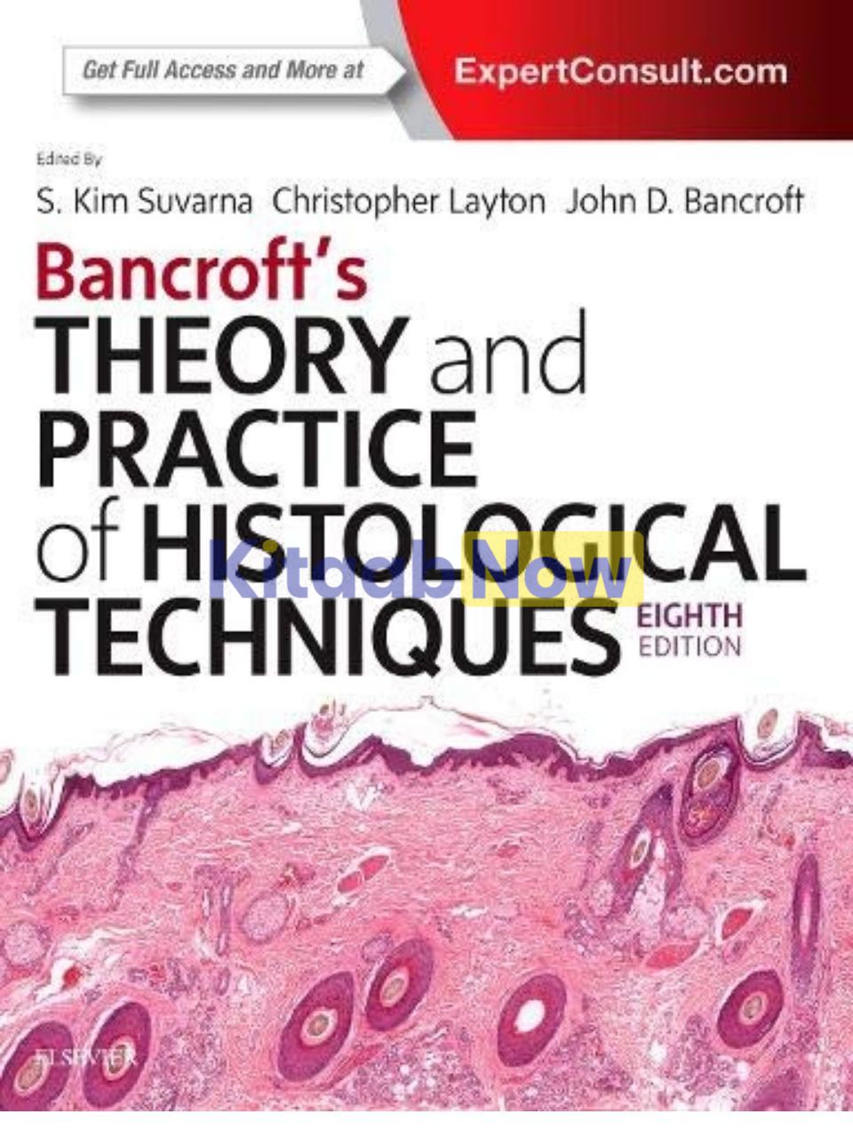
Bancroft’s Theory and Practice of Histological Techniques (8th Edition)
- Authors: Kim S Suvarna, Christopher Layton, John D. Bancroft
- ISBN: 9780702068645
- Publisher: Elsevier Science Publishing,
- Edition: 8th
- Publication Date: May 02, 2018
- Format: Paperback – 584 pages
- Language: English
Description
For 40 years, Bancroft’s Theory and Practice of Histological Techniques has established itself as the standard reference for histotechnologists and laboratory scientists, as well as histopathologists. With coverage of the full range of histological techniques used in medical laboratories and pathology departments, it provides a strong foundation in all aspects of histological technology – from basic methods of section preparation and staining, to advanced diagnostic techniques such as immunocytochemistry and molecular testing. This revised and updated 8th Edition by Kim S. Suvarna, Christopher Layton, and John D. Bancroft is a one-stop reference for all those involved with histological preparations and applications, from student to highly advanced laboratory professional.
New to this Edition
- Features new content on automation (computer logging and tracking, sub-sampling and archiving of samples), digital scanning techniques for slides used in remote/telepathology, and specialist molecular techniques.
- Provides superb visual guidance through the use of vivid color illustrations, including additional illustrations of diagnostic modalities and techniques.
- Contains more summary tables, charts, and boxes throughout for quick reference.
- Expert Consult™ eBook version included with purchase. This enhanced eBook experience allows you to search all of the text, figures, and references from the book on a variety of devices.
Key Features
- Presents a thorough, up-to-date, and detailed approach to the theory and protocols for preparing cells and tissues for laboratory evaluation, covering topics ranging from basic to advanced
Table of Contents
- Pathology Laboratory Management
- Chemical Safety in the Laboratory
- Light Microscopy
- Fixation of Tissues
- The Gross Room
- Tissue Processing
- Microtomy
- Resin Techniques
- Theory of Histological Staining
- The Hematoxylins and Eosin
- Automation in the Histology Department
- Connective and other Mesenchymal tissues with their stains
- Carbohydrates
- Pigments and minerals
- Amyloid
- Traditional stains and modern techniques for demonstrating Micro-organisms in Histology
- Bone
- Neuropathology and Muscle Biopsy Techniques
- Immunohistochemical and Immunofluorescent Techniques
- Molecular Pathology
- Transmission Electron Microscopy
- Digital Pathology
- Appendices
- Diagnostic Appendices
- Appendix I Traditional Methods
- Appendix II Tissue Microarray
- Appendix III Applications of Immunohistochemistry
- Technical Appendices
- Appendix IV Measurement Units
- Appendix V Preparation of Solutions
- Appendix VI Buffer Solutions
- Appendix VII Solubility of Some Common Reagents and Dyes
- Appendix VIII Mounting Media and Slide Coatings
Author Biography
Kim S Suvarna, MBBS, BSc, FRCP, FRCPath, Department of Pathology, Northern General Hospital, Sheffield;
Christopher Layton, PhD, Specialist Section Lead in Specimen Dissection, Histopathology Department, Northern General Hospital, Sheffield, UK
John D. Bancroft, Formerly Pathology Directorate and Business Manager, Queen’s Medical Center, Nottingham, UK
Additional information
| Weight | 1.470 kg |
|---|
Only logged in customers who have purchased this product may leave a review.
Related Products




Reviews
There are no reviews yet.